Restrictive Lung Disease: An Overview
New Title

Abstract
Restrictive lung disease (RLD) encompasses a diverse group of respiratory disorders characterized by reduced lung expansion, leading to decreased lung volume and increased effort in breathing. This paper aims to provide a comprehensive overview of RLD, including its etiology, pathophysiology, clinical manifestations, diagnostic approaches, and treatment options. Emphasis is placed on understanding the underlying mechanisms, the impact on patients’ quality of life, and current research trends in the field.
Introduction
Restrictive lung disease (RLD) is a category of pulmonary disorders that impede the lungs' ability to expand fully during inspiration, resulting in reduced total lung capacity (TLC). Unlike obstructive lung diseases, where airflow is restricted due to narrowed airways, RLD primarily involves the lung parenchyma, pleura, chest wall, or neuromuscular apparatus. This paper explores the various causes, diagnostic methods, clinical management, and emerging research on RLD.
Etiology
Intrinsic Causes
Intrinsic causes of RLD involve diseases that affect the lung parenchyma, leading to fibrosis, inflammation, or scarring. Common intrinsic causes include:
- Idiopathic Pulmonary Fibrosis (IPF): A chronic, progressive fibrotic disorder of unknown etiology.
- Sarcoidosis: A multisystem inflammatory disease characterized by granuloma formation.
- Pneumoconiosis: Lung diseases caused by the inhalation of dust, often in occupational settings (e.g., asbestosis, silicosis).
- Hypersensitivity Pneumonitis: An immune-mediated condition resulting from inhalation of environmental antigens.
Extrinsic Causes
Extrinsic causes are related to conditions that restrict lung expansion from outside the lung tissue, such as:
- Chest Wall Disorders: Conditions like kyphoscoliosis or severe obesity that mechanically limit lung expansion.
- Pleural Diseases: Disorders like pleural effusion, pleural fibrosis, or pneumothorax.
- Neuromuscular Diseases: Conditions such as amyotrophic lateral sclerosis (ALS) or muscular dystrophies that impair respiratory muscle function.
Pathophysiology
The pathophysiology of RLD involves reduced compliance of the lung tissue or the structures surrounding the lungs. This decreased compliance results in increased work of breathing and reduced lung volumes, particularly the TLC and functional residual capacity (FRC). In intrinsic RLD, alveolar inflammation and fibrosis disrupt the lung architecture, leading to impaired gas exchange. In extrinsic RLD, mechanical factors limit lung expansion.
Clinical Manifestations
Patients with RLD typically present with:
- Dyspnea: Shortness of breath, particularly during exertion.
- Cough: Often dry and persistent.
- Fatigue: Resulting from increased work of breathing and hypoxemia.
- Chest Discomfort: Occasionally reported, particularly in cases involving pleural disease.
Diagnostic Approaches
Pulmonary Function Tests (PFTs)
PFTs are crucial for diagnosing RLD. Key findings include:
- Reduced TLC: Indicative of restricted lung expansion.
- Normal or Increased FEV1/FVC Ratio: Unlike obstructive lung diseases, the FEV1/FVC ratio is typically normal or increased due to proportionally reduced FVC.
Imaging Studies
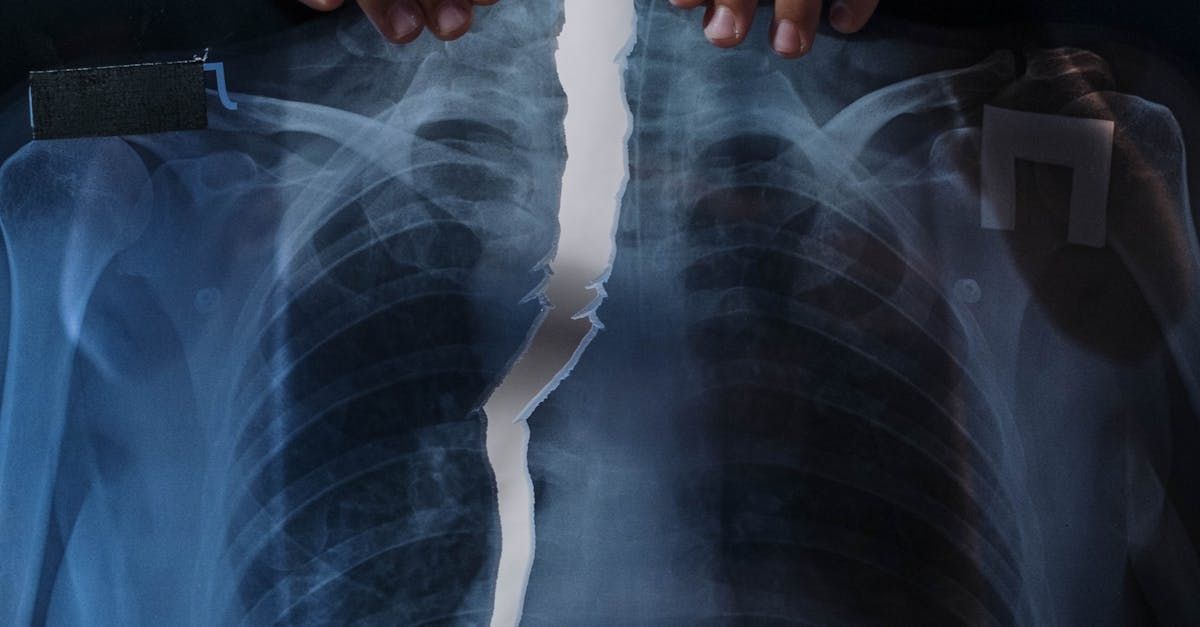
- Chest X-ray: Can reveal lung parenchymal changes, chest wall abnormalities, or pleural disease.
- High-Resolution Computed Tomography (HRCT): Provides detailed images of lung parenchyma, aiding in the diagnosis of interstitial lung diseases.
Additional Tests
- Arterial Blood Gases (ABGs): Assess oxygenation and ventilation status.
- Biopsy: In cases where the diagnosis is unclear, lung biopsy may be performed to identify specific histopathological features.
Treatment Options
Pharmacologic Therapy
- Antifibrotic Agents: For IPF, drugs like pirfenidone and nintedanib can slow disease progression.
- Corticosteroids and Immunosuppressants: Used in inflammatory causes of RLD, such as sarcoidosis or hypersensitivity pneumonitis.
Non-Pharmacologic Therapy
- Pulmonary Rehabilitation: Exercise training and education to improve physical function and quality of life.
- Oxygen Therapy: For patients with significant hypoxemia.
- Mechanical Ventilation: In advanced neuromuscular diseases, non-invasive or invasive ventilation may be required.
Surgical Interventions
- Lung Transplantation: Considered for patients with end-stage RLD refractory to medical therapy.
Emerging Research
Recent research in RLD focuses on identifying novel biomarkers for early diagnosis, understanding the genetic basis of fibrotic lung diseases, and developing targeted therapies to halt or reverse lung fibrosis. Advances in imaging techniques and computational modeling are also enhancing our understanding of disease progression and treatment response.
Conclusion
Restrictive lung disease encompasses a broad spectrum of conditions that significantly impact patients’ respiratory function and quality of life. Early diagnosis and comprehensive management are essential to improving outcomes. Continued research is critical to uncovering the underlying mechanisms and developing effective therapies for these challenging disorders.
References
- Raghu, G., et al. (2018). Diagnosis of Idiopathic Pulmonary Fibrosis: An Official ATS/ERS/JRS/ALAT Clinical Practice Guideline. American Journal of Respiratory and Critical Care Medicine, 198(5), e44-e68.
- Drent, M., et al. (2020). Sarcoidosis: Diagnosis and Management. European Respiratory Review, 29(156), 200002.
- Hunninghake, G. W., & Lynch, D. A. (2017). Approach to the Adult with Interstitial Lung Disease: An Update. Annals of the American Thoracic Society, 14(Supplement_2), S108-S114.
- Flaherty, K. R., et al. (2019). Nintedanib in Progressive Fibrosing Interstitial Lung Diseases. New England Journal of Medicine, 381(18), 1718-1727.
- Ley, B., Collard, H. R., & King, T. E. (2011). Clinical Course and Prediction of Survival in Idiopathic Pulmonary Fibrosis. American Journal of Respiratory and Critical Care Medicine, 183(4), 431-440.
- Holland, A. E., et al. (2014). Pulmonary Rehabilitation for Interstitial Lung Disease. Cochrane Database of Systematic Reviews, (10), CD006322.
- Lederer, D. J., & Martinez, F. J. (2018). Idiopathic Pulmonary Fibrosis. New England Journal of Medicine, 378(19), 1811-1823.
- Nathan, S. D., et al. (2016). Efficacy of Pulmonary Rehabilitation in Patients with Idiopathic Pulmonary Fibrosis: Results of the Pulmonary Rehabilitation in Idiopathic Pulmonary Fibrosis (PRIDE) Study. Chest, 150(4), 759-768.
This post provides a comprehensive overview of restrictive lung disease, highlighting key aspects such as etiology, pathophysiology, clinical manifestations, diagnostic approaches, and treatment options. The references cited support the information provided and guide further reading on this topic.
